Metabolomics
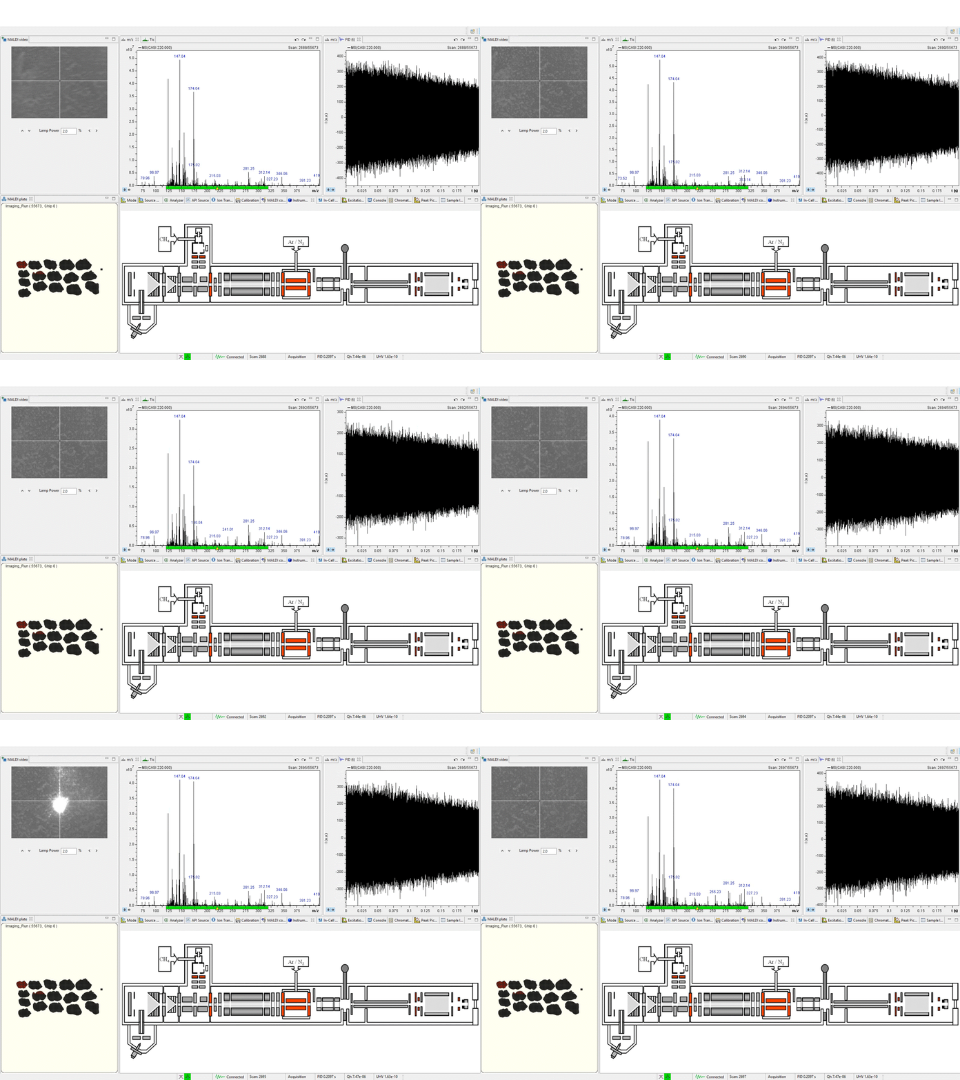
Cellular metabolism is dependent on enzyme-catalyzed changes to carbohydrates, lipids, and proteins. Utilizing the power of cutting-edge mass spectrometry, it is possible to spatially pinpoint within a diseased tissue where metabolic aberrations occur. The SMIL frequently uses Matrix Assisted Laser Desorption/Ionization (MALDI) paired with either Time of Flight or Molecular Resonance mass spectrometry to broadly identify metabolic molecule concentration and location within a given tissue. The applications of these resources are vast, and our current work focuses on several types of brain cancers, breast cancer, and prostate cancer.
Pharmacometabolomics

Our research focuses on the development and implementation of integrated biomolecular and drug imaging of tissue specimens. By tuning our instruments specifically to a drug molecule of interest, it is possible to assess drug delivery within a tissue. This research is central to combatting glioblastoma and other brain cancers, as assessing drug delivery through the blood-brain barrier is key for the identification of novel treatments.

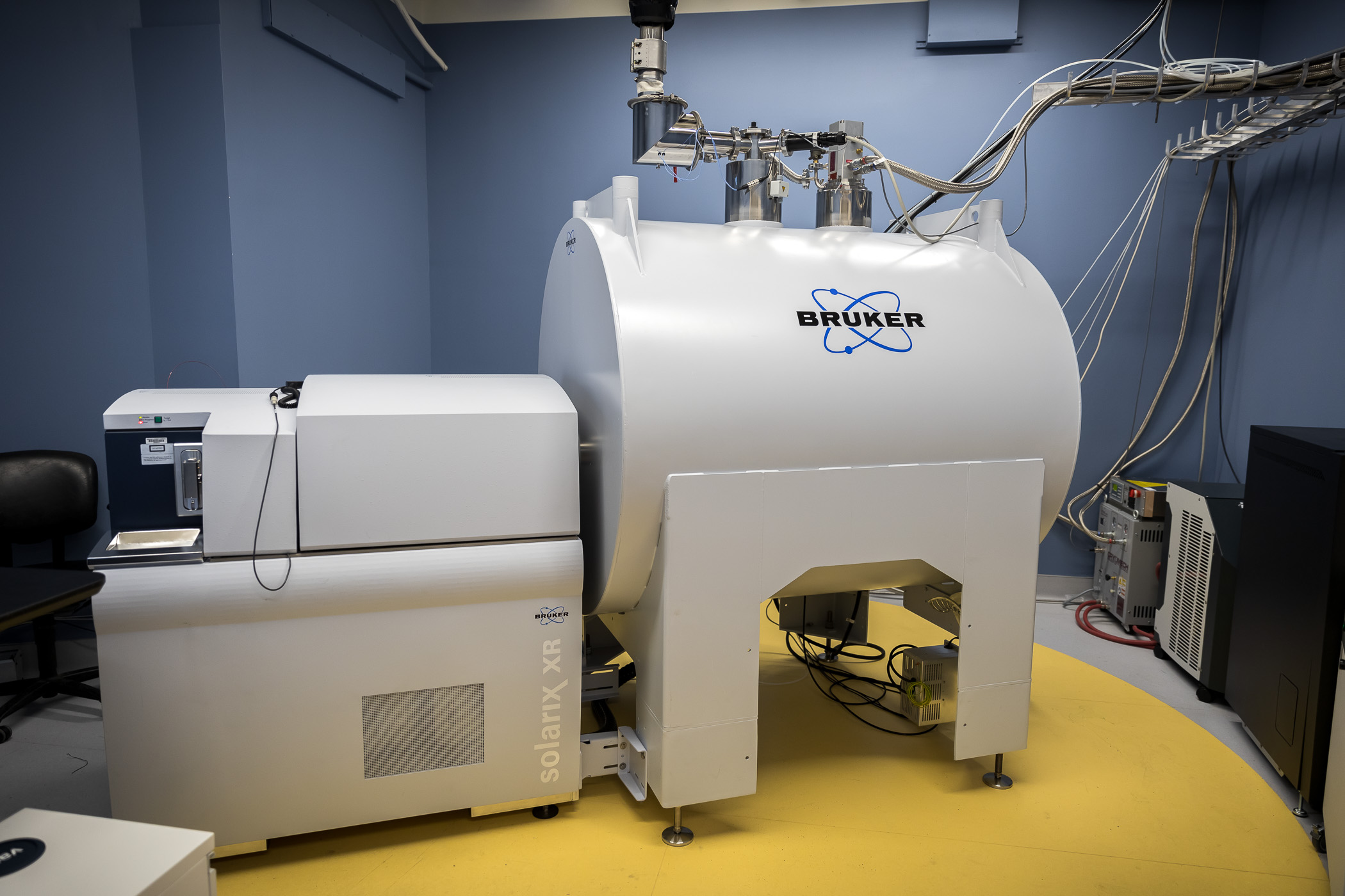
Our 15Tesla SolariX XR Magnetic Resonance Mass Spectrometer by Bruker Daltronics allows for Fourier-Transform Ion Cylcotron Resonance mass spectrometry. Awarded through the Massachusetts Life Sciences Center, the SMIL leverages our “FT’s” precision and accuracy in molecular identification to contest cancer and disease. By imaging drugs and metabolic profiles from pre-clinical animal models and clinical trial specimens, we hope to discover answers to the unmet need for therapeutics for brain tumors.
Developing and Validating Real-Time Mass Spectrometry Approaches

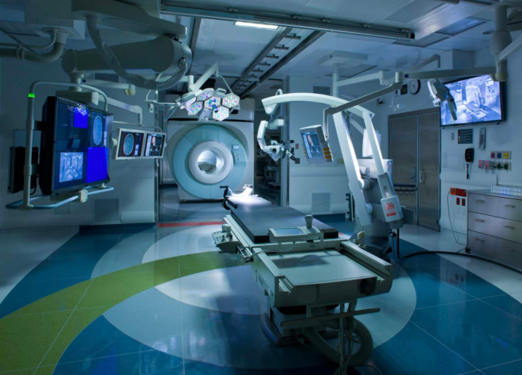
Timing is everything, and implementing fast, reliable measurements on surgical samples is critical in surgical decision-making. Here at SMIL, we are actively investigating and providing support through mass spectrometry imaging to improve outcomes in the operating room for patients of today and of tomorrow. We have installed and operate a mass spectrometer in the Advanced Multi-Modality Image Guided Operating suite (AMIGO) at Brigham and Women’s Hospital to provide real-time surgical guidance support to our neurosurgeons. It is our goal to enable surgeons and oncologists to tailor treatment from the time of surgery, support the development of new therapeutics, and allow precision cancer care using molecular imaging with mass spectrometry approaches.
Broad Applications

Not limited to oncology, our research collaborations widen our scope for harnessing MSI for other research disciplines. Particularly in Neuroscience and Developmental Biology, our collaborations expand our research efforts for discovery in improved epilepsy treatments (Yellen Lab) and in human diseases of the musculo-skeletal axis (Pourquié Lab).


